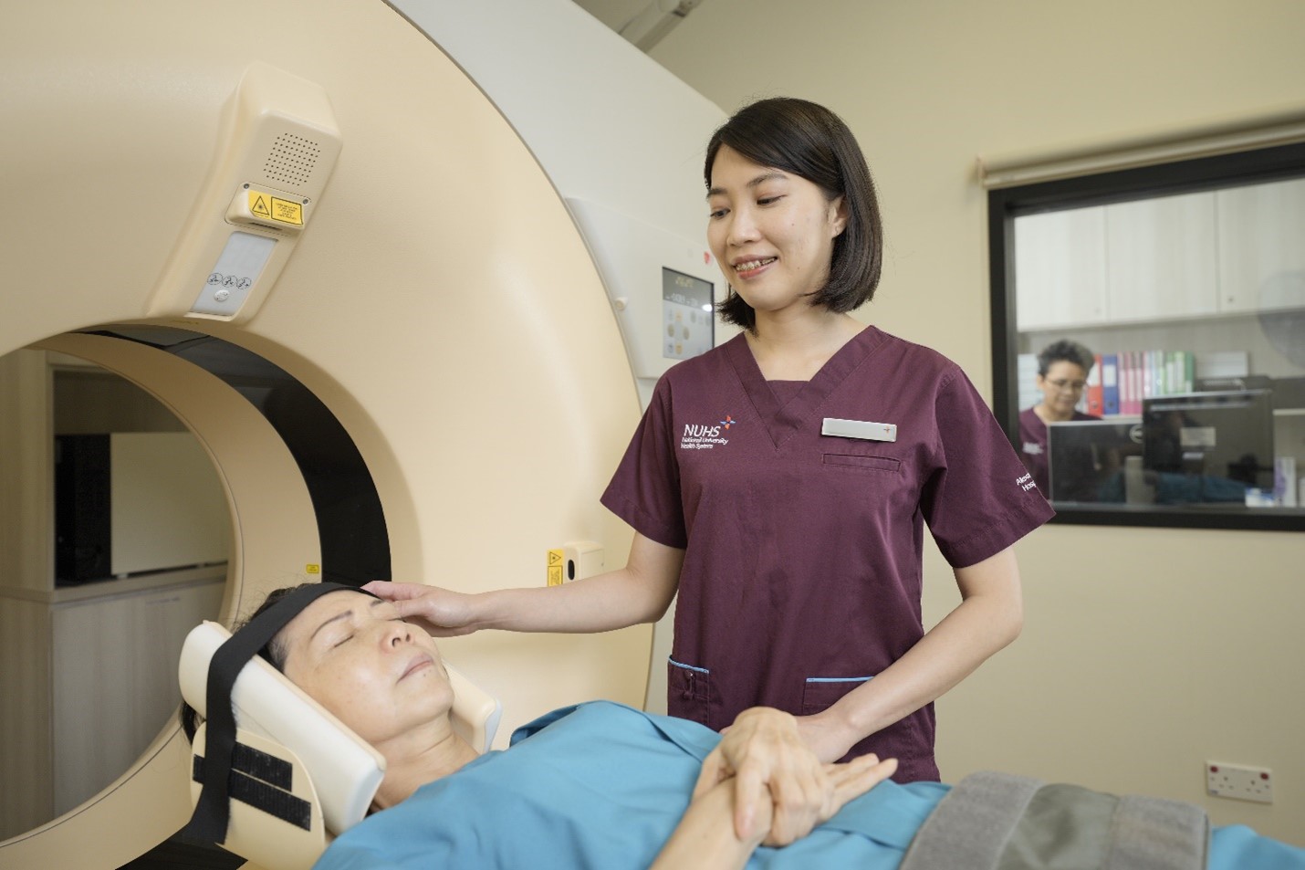
Radiography/Diagnostic Imaging's core objective is to deliver comprehensive multidisciplinary medical imaging services for the patients. Radiographers in the department utilise a complete range of diagnostic and interventional imaging modalities, along with PACS (Picture Archiving and Communications), RIS (Radiology Information System), and the hospital NGEMR system. We work closely with our Radiologist, Imaging Informatics and Admin colleagues to provide patient care through imaging examinations.
Such imaging modalities included X-ray, sonography, mammography, computed tomography (CT), magnetic resonance imaging (MRI), bone mineral densitometry (using DEXA scanner), fluoroscopy as well as angiography system inside our interventional suite etc.
'Coming together is a beginning, staying together is progress, and working together is success.' – Henry Ford.
I'm glad to have been here since Alex started its operations back in mid-2018, experiencing the challenges, growth and progress that our team has achieved throughout the years, including the pandemic journey. Focusing on 'working together' as highlighted by Henry Ford, has been the critical element in our daily work, and with the new campus and team expansion coming soon, I am excited with the possibilities and developments that we can achieved in future both individual and as a team.
I am a radiographer specializing in sonography. I deliver scans and direct care for patients by using the ultrasound machine. The day starts with performing quality assurance checks for machines before they are utilised for the examinations. Subsequently, once a patient has arrived and an identifiers check is done, the ultrasound scan will be performed for patient. Further instructions will then be given to the patient once the scan is done, and the images are reviewed. Finally, preliminary report will be done for each scan and passed over to the Radiologist for reporting.
Ultrasound scan is a real-time examination with no radiation. It plays a critical role in not only health screening but also in detecting abnormalities and providing diagnosis. Ultrasound scan is constantly required for further characterisation of lesion incidentally found in CT (Computed Tomography) or MRI (Magnetic Resonance Imaging) scan, as ultrasound provide detailed information such as lesion's contour, composition, vascularity, etc. The information helps our doctors to make accurate diagnosis and clinical decisions on whether certain treatment or intervention is needed.